

Diagnostic Tests
Imaging Tests
A patient’s doctor will most likely order imaging tests to help find areas of the body where there may be cancer, to learn how far the cancer has spread, and later on to check how well the treatment is working. Most of these tests are painless and no anesthetic is required.
Choose from the menu or scroll down to read about imaging tests:
X-Ray
X-rays use radiation to take pictures of areas inside the body. The amount of radiation used in most diagnostic tests is so small that it poses little risk to the patient. Findings on a chest x-ray may indicate whether the disease is bulky (large tumors or lymph nodes, usually resistant to conventional therapy).
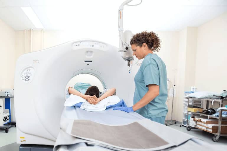
Computer Tomography (CT) Scan
A CT scan takes x-rays from many different angles around the body. A computer combines the pictures obtained from these different angles to give a detailed image of organs inside the body. Patients with lymphoma often have CT scans of the neck, chest, abdomen, and pelvis to find out how many lymph nodes are involved, how large they are, and whether internal organs are affected by the disease.
The amount of radiation exposure during a CT scan varies depending on the area scanned. Most CT scans confer little risk to the patient, although CT scans of the abdomen and pelvis do add a moderate amount of risk (1 in 500-1,000) to the general lifetime risk of cancer.
Before a CT scan, the patient may be asked to drink a contrast liquid and/or get an intravenous injection of a contrast dye that will more clearly outline abnormal areas that may be present in the body.
Magnetic Resonance Imaging (MRI)
Like a CT scan, an MRI takes images from different angles around the body, but an MRI does not use x-rays like a CT scan; instead it uses magnets and radiofrequency waves. Therefore, an MRI confers no risk of radiation-induced cancer.
An MRI can provide important information about tissues and organs, particularly the nervous system, that is not available from other imaging techniques. Because this testing technique works well to get clear images of the bones, brain, and spinal cord, an MRI may be ordered if a doctor wants to see whether the lymphoma has spread in these areas.
Positron Emission Tomography (PET) Scan
To perform the test, radioactive fluorodeoxyglucose (a type of sugar) is first injected into the body. A positron camera is then used to detect the radioactivity and produce cross-sectional images of the body. The amount of radiation used for a PET scan is low and poses little risk to the patient.
PET scans help determine how much disease is present (staging) and how well it is responding to treatment. While CT scans show the size of a lymph node, PET scans show if the lymph node is active (still has disease). CT and PET scans are now combined into one test (PET/CT).
Multi-Gated Acquisition (MUGA) Scan
A MUGA scan is an imaging test that looks at how well the heart muscle is working. A MUGA scan is done to make sure that the patient’s body can withstand treatment with certain lymphoma drugs that may damage the heart in rare cases. MUGA scans may be done when patients are resting or exercising, depending on what their doctor wants to assess.
A doctor will most likely order a MUGA scan if he or she is considering treating a patient with the drug doxorubicin (Adriamycin). This test is needed to make sure that the heart is functioning normally because doxorubicin may be associated with cardiac toxicity. A two-dimensional echocardiogram (ECHO) is sometimes used instead of the MUGA scan to test heart function.
Also Learn About: